


Importance of Early Brain Tumor Detection
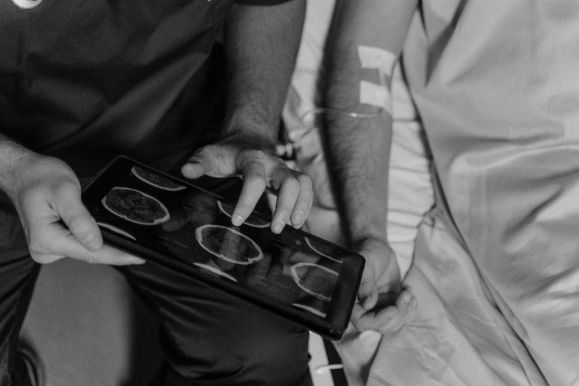
Early detection of brain tumors is a key aspect of treatment for brain tumors. Brain tumors, whether benign or malignant, can lead to a wide range of neurological symptoms and complications. Detecting them at an early stage can significantly increase the chances of successful treatment and patient survival. Recent advances in brain tumor imaging technologies have paved the way for more precise, non-invasive, and timely diagnosis.
Advanced Imaging Techniques
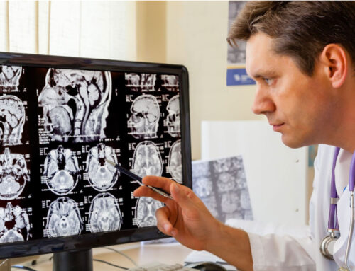
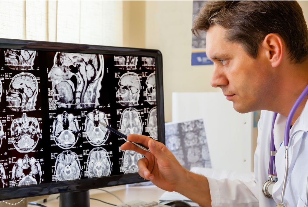
Magnetic Resonance Imaging (MRI)
Recent enhancements in MRI technology have allowed for more detailed and high-resolution imaging of brain tumors. Functional MRI (fMRI) can assess brain activity by measuring changes in blood flow, helping to locate areas affected by tumors. Diffusion tensor imaging (DTI) can also map the white matter tracts of the brain, providing very important information for surgical planning.
Positron Emission Tomography (PET)
PET scans combined with newer radiotracers have improved the ability to detect and characterize brain tumors. These scans can not only identify the presence of tumors but also assess their metabolic activity, aiding in the differentiation between benign and malignant lesions.
Computed Tomography (CT)
Advancements in CT imaging have resulted in faster and more accurate scans. Dual-energy CT can provide valuable information about tissue composition, helping in the characterization of brain lesions. The use of contrast agents in CT angiography also enables the visualization of blood vessels within and around tumors.
Functional Imaging
One of the most significant recent innovations in brain tumor detection is the incorporation of functional imaging techniques. These approaches provide insights into the tumor’s biological behavior, helping clinicians make informed decisions about treatment strategies.
1. Spectroscopy Imaging
Spectroscopy imaging measures the chemical composition of tissues, allowing for the identification of specific metabolites associated with brain tumors. By analyzing these metabolic profiles, clinicians can distinguish between tumor types and assess treatment response.
2. Perfusion Imaging
Perfusion imaging evaluates blood flow within and around brain tumors. It provides valuable information about tumor vascularity, which can aid in grading and treatment planning. Techniques like dynamic susceptibility contrast (DSC) and arterial spin labeling (ASL) have advanced perfusion imaging capabilities.
3. Functional MRI (fMRI)
As mentioned earlier, fMRI assesses brain activity. It is particularly useful in identifying eloquent brain regions that should be spared during surgery. Additionally, resting-state fMRI can reveal alterations in brain connectivity caused by the presence of a tumor.
Advanced Software and Artificial Intelligence
Recent developments in imaging software and artificial intelligence (AI) have transformed the interpretation of brain scans. AI algorithms can analyze large datasets of imaging studies, identifying subtle patterns and anomalies that may escape human detection. This technology can aid radiologists in making faster and more accurate diagnoses, ultimately benefiting patients by reducing the time to treatment initiation.
Radiomics and Machine Learning
Radiomics is an emerging field that extracts quantitative data from medical images. By analyzing thousands of features within an image, radiomics can provide insights into tumor heterogeneity, growth patterns, and treatment response. Machine learning algorithms process this radiomic data, enabling predictive modeling and personalized treatment recommendations.
Advanced Imaging for Intraoperative Guidance
Intraoperative imaging has also seen significant advancements, particularly in brain tumor surgery. Techniques like intraoperative MRI (iMRI) and intraoperative CT (iCT) allow surgeons to obtain real-time images during the procedure. This capability enables precise tumor resection while minimizing damage to healthy brain tissue.
Advanced Imaging for Targeted Therapy
Targeted therapies for brain tumors have become more effective due to recent imaging innovations. Molecular imaging techniques can visualize specific molecular markers on tumor cells, aiding in the selection of appropriate targeted therapies. This personalized approach enhances treatment outcomes while reducing potential side effects.
Conclusion
Recent imaging advances have transformed the landscape of early brain tumor detection and diagnosis. These innovations, ranging from advanced imaging techniques and functional imaging to artificial intelligence and intraoperative guidance, empower healthcare professionals to provide more accurate and timely care to patients. By staying informed about these breakthroughs, individuals can make more informed decisions about their healthcare and contribute to the ongoing progress in the field of brain tumor diagnosis and treatment.
As research and technology continue to evolve, the future holds even more promise for improving the early detection and management of brain tumors. For patients in Orange County and beyond, these advancements offer hope for earlier intervention, better treatment outcomes, and ultimately, an improved quality of life.


